
 Buy $FLYMIND
FLYMIND
In 2024 the FlyWire consortium published the first complete wiring diagram of an adult animal's brain: every neuron of a fruit fly and every synapse between them, traced from electron microscopy. FlyMind is a specimen viewer for that map, 139,255 neurons and about 50 million connections rendered from the published counts, with $FLYMIND on Robinhood Chain as its attestation.
Anatomical specimen
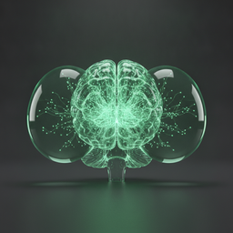
One point for ten neurons. The shape is a schematic model built from the published region layout, not the dataset itself; the counts on the right are FlyWire's. Drag to rotate.
What does a whole brain look like when every wire is known?
For a century neuroscience worked from samples: a few neurons stained, a few circuits traced. In 2024 a consortium led from Princeton, with hundreds of labs and volunteer proofreaders, finished the map of an entire adult brain, that of a female fruit fly. The raw material was a brain sliced into thousands of sections and imaged with electron microscopes; the segmentation was done by AI and corrected by people, synapse by synapse.
A whole brain sliced and photographed
The FAFB volume: one adult female fly brain cut into thousands of ultrathin sections and imaged with serial-section electron microscopy at nanometre resolution, published in 2018 as an open dataset.
Machine learning draws the neurons
Convolutional networks traced every neuron through the image stack. FlyWire opened the result to the community as a proofreading platform, where people fix merges and splits the model got wrong.
The larval brain, complete
A related effort finished the connectome of a fly larva first: 3,016 neurons and about 548,000 synapses, published in 2023 as the first full brain wiring diagram of an insect at any stage.
139,255 neurons, every synapse
The adult connectome and its cell-type atlas appeared together in Nature: 139,255 neurons, about 50 million synapses, 8,453 cell types, released openly for anyone to query.
How a fly sees a threat and leaves
The connectome makes it possible to follow a signal across the whole brain instead of stopping at a region boundary. The route below is the anatomy the map traces for vision.
Photoreceptors
Each of the compound eye's units carries photoreceptor cells that turn light into membrane voltage.
Optic lobe layers
The optic lobes, a large share of all neurons in the brain, sort the image by contrast, motion and direction in repeated columns.
Motion detectors
Wide-field neurons integrate motion across the visual field; a looming shape reads as an approaching object.
Down to the body
Descending neurons carry the decision from the brain to the ventral nerve cord, where the legs and wings are driven.
Slice inspector
Move through the specimen front to back. Each slice is a cross-section of the schematic model above, so what you see is where the regions sit, not a micrograph.
Staining mode
Anterior to posterior
The specimen and its sources
Drosophila melanogaster, adult female
The FAFB brain: one individual, imaged whole. The connectome describes this one fly; a second brain, a male, is being mapped to compare.
Serial-section EM, AI segmentation, human proofreading
Sections at nanometre resolution, neurons traced by convolutional networks, corrections by hundreds of proofreaders including volunteers, synapses detected automatically.
Open data
The connectome is public and queryable. FlyMind draws only the published counts and region layout; nothing here is measured by us.
$FLYMIND on Robinhood Chain
$FLYMIND is an ordinary Pons coin. It has no claim on the data, the labs, or the fly. What it does is give this specimen viewer an address on Robinhood Chain: the contract below is the token, the numbers above are the consortium's, and the two are kept apart on purpose.